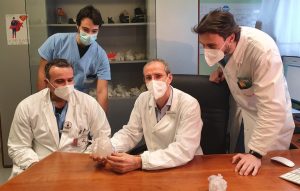
The press release received from the Hospital/University of Padua contained several medical technicalities, but the central concept was clear: the Department of Pediatric Surgery of Padua now uses 3D modeling to train surgeons, dealing with complex and always different cases.
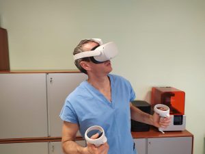 Virtual Reality has become an essential resource for the team of Prof Vladimiro Vida, Director of Pediatric Cardiac Surgery: 3D modeling allows to “navigate”, but also to manipulate and interact with objects, in this case the hearts of patients, expanding the possibilities both to study a wide range of anatomical and pathophysiological variables, and to investigate existing cases for future interventions.
The potentialities of this innovative technology are many and are all aimed at improving the quality of treatment offered to patients, in three main areas: prenatal counseling to families, to define treatments for the newborn, or in the preoperative phase, but also to better plan the surgery. Printed models allow to simulate surgical procedures to optimize time and materials, and to improve the understanding of the specific case through the study of models, especially in congenital heart disease.
The Azienda Ospedale/University of Padua has at its disposal technologies that, starting from different types of traditional cardiac “imaging”, such as computerized axial tomography (CT), nuclear magnetic resonance (NMR) and fetal ultrasound, have the possibility of achieving a three-dimensional reconstruction of the cardiac organ affected by malformations; thanks to 3D printing it is also possible to obtain a reproduction of the cardiac organ that is as faithful as possible to the patient’s real organ.
The press release closes with a note: the experience of surgeons on real cases remains irreplaceable, and this is easy to understand, but this use of virtual and immersive reality is a great help, probably irreplaceable too, in of today’s surgeons education.